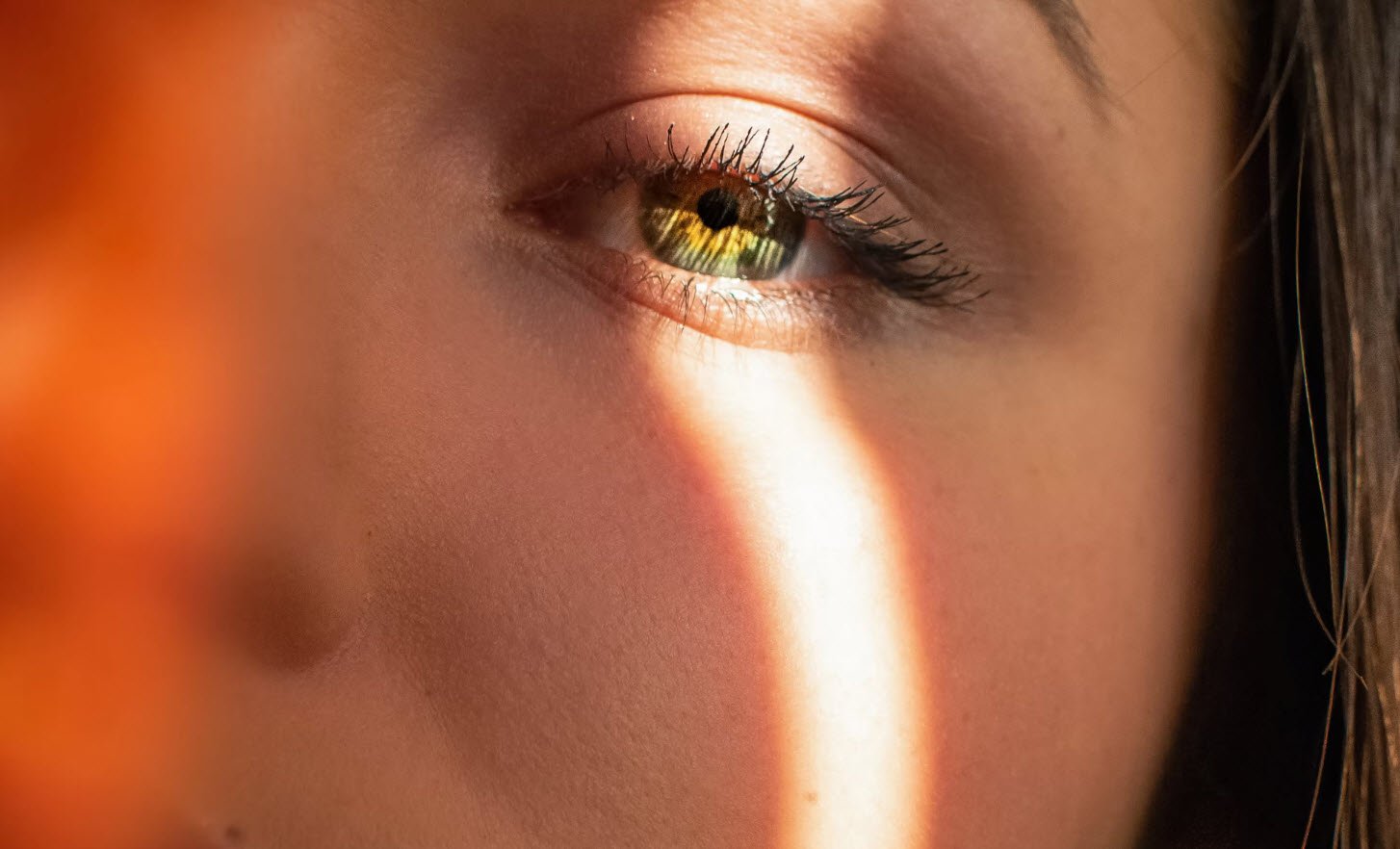
A number of things can go wrong in any of the structures of the eye, causing visual impairment and blindness. Many of these deteriorations are due to ageing.
Also Read: 10 Proven Tips for Fighting Acne And Get Clear Skin
The main visual disorders are described below.
- Glaucoma
- Cataract
- Age-related macular degeneration
- Diabetic retinopathy
- Retinitis pigmentosa
- Pterygium
- Ocular surface neoplasia
1. Glaucoma
Glaucoma is a group of diseases of the optic nerve that result in loss of vision or blindness. The optic nerve is gradually destroyed, usually due to excessively high intraocular pressure, known as ocular hypertension (OHT).
Open-angle glaucoma (OAG) is the most common form of glaucoma. It occurs when the fluid in the anterior chamber reaches the angle between the cornea and iris, and passes too slowly through the meshwork drain. As the fluid builds up, the pressure inside the eye rises to a level that may damage the optic nerve.
Different people’s eyes can withstand different levels of intraocular pressure, and a diagnosis of glaucoma requires damage to the optic nerve rather than OHT alone.
Glaucoma can also develop without increased eye pressure (called low-tension or normaltension glaucoma). Causes include poor blood supply to the optic nerve fibres, a weakness in the optic nerve structure or a problem with the optic nerve fibres.
2. Cataract
Cataracts are a clouding of the lens, resulting in a blurred image. The lens is made of mostly water and protein, arranged in a way that keeps the lens clear and allows light to pass through.
As we age, some of the protein may clump together and start to cloud a small area of the lens, resulting in a cataract that reduces the amount of light reaching the retina. Over time, the cataract may slowly grow larger and cloud more of the lens, making vision gradually duller or more blurred.
There are three main types of cataract:
- nuclear cataracts are the most common type, so named because they occur in the centre of the lens
- cortical cataracts start in the cortex (periphery) of the lens and gradually extend to the centre of the lens
- subcapsular cataracts are those in which the opacities are concentrated beneath or within the capsule of the lens.
Cataract development is part of the ageing process. Hence, although lifestyle changes may reduce the rate of development for some people, cataracts cannot be prevented. However, surgery can correct the condition.
3. Age-related macular degeneration
In AMD, clumps of yellowish material (known as drusen) gradually accumulate within and beneath the RPE. The RPE cells may die and therefore no longer support the photoreceptors, which then cannot function, resulting in loss of vision in that part of the retina.
If the photoreceptors in the macula are affected, this can seriously impair fine visual skills; this situation is sometimes referred to as ‘dry’ macular degeneration (or central, geographic atrophy), and is the most common form of AMD.
In about 10% of cases, dry AMD progresses to a more advanced and damaging form of the eye disease known as wet macular degeneration (or neovascular AMD). New blood vessels grow beneath the retina to supply more nutrients and oxygen to the retina.
Suggested Read: 3 Tips on how to remove Acne Scars and Blemishes
However, often these blood vessels are in areas where they are not supposed to be; for example, in the macula. Bleeding, leaking and scarring from these blood vessels affect the photoreceptors and impair vision.
4. Diabetic retinopathy
Diabetic retinopathy is a common complication of diabetes. Poor glucose control during diabetes affects the tiny blood vessels of the retina. The arteries become weakened and leak blood, leading to swelling or oedema in the retina and decreased vision.
As diabetes progresses, circulation problems can also deprive the retina of oxygen. New, fragile, vessels develop; however, these are susceptible to bleeding and the blood may leak into the retina and vitreous, causing spots or floaters, along with decreased vision. If abnormal blood vessel growth continues, scar tissue formation may cause retinal detachment and glaucoma may also develop.
5. Retinitis pigmentosa
Retinitis pigmentosa refers to the group of genetic eye diseases that causes degeneration of photoreceptor cells in the retina. As these cells degenerate and die, patients experience progressive vision loss. The condition is diagnosed by using electroretinography to document progressive loss in photoreceptor function.
6. Pterygium
A pterygium is a noncancerous growth of the clear, thin tissue that overlies the white part of the eye (the conjunctiva). The condition may affect one or both eyes. Although the cause of pterygia is unknown, people who work outdoors and are excessively exposed to sunlight and wind more frequently develop pterygia than people who work indoors.
Pterygia present as a painless, raised area of white tissues, with blood vessels on the inner or outer edge of the cornea. No treatment is usually required unless the pterygia begins to obstruct vision, and surgical removal of the pterygium usually has good results.
7. Ocular surface neoplasia
Ocular surface neoplasia is term used to describe various dysplasias, carcinoma in situ and squamous cell carcinoma of the ocular surface epithelium (conjunctiva and cornea).
You may also like:- Top 10 Tips for Effectively Whitening Your Teeth
- 8 Essential Tips for Healthy and Shining Skin
- 7 Tips for Boys on Reducing Acne
- 7 Tips for Maintaining Acne-Free Skin
- 8 Common Hair Loss Myths You Need To Know
- The Two-Step Skin Care Routine Everyone Should Follow
- Effective Ways to Fade Unsightly Acne Scars
- 3 Healthy Anti-Aging Skin Care Tips for Youthful Radiance
- Top 10 Skin Care Tips for a Radiant Complexion
- Hair Replacement – 10 Useful Tips for a Confident Decision


